Περιγραφή
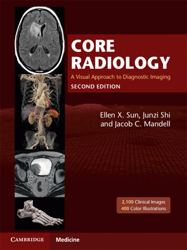
Embodying the principle of ‘everything you need but still easy to read’, this fully updated edition of Core Radiology is an indispensable aid for learning the fundamentals of radiology and preparing for the American Board of Radiology Core exam. Containing over 2,100 clinical radiological images with full explanatory captions and color-coded annotations, streamlined formatting ensures readers can follow discussion points effortlessly. Bullet pointed text concentrates on essential concepts, with text boxes, tables and over 400 color illustrations supporting readers’ understanding of complex anatomic topics. Real-world examples are presented for the readers, encompassing the vast majority of entitles likely encountered in board exams and clinical practice. Divided into two volumes, this edition is more manageable whilst remaining comprehensive in its coverage of topics, including expanded pediatric cardiac surgery descriptions, updated brain tumor classifications, and non-invasive vascular imaging. Highly accessible and informative, this is the go-to introductory textbook for radiology residents worldwide.
- Fully updated to include state of the art and emerging topics such as expanded pediatric cardiac surgery descriptions, updated brain tumor definitions and non-invasive vascular imaging
- Streamlined text design and formatting ensures that images are always aligned with the relevant text, meaning readers can effortlessly follow the discussion
- Contains over 2,100 clinical radiological images with full explanatory captions and color-coded annotations that are consistent throughout the book, allowing readers to identify common features between cases
- Over 400 high-quality, full-color illustrations engage readers in the subject matter and ensure they can easily grasp complex anatomic topics
- Divided into two volumes, the book is more manageable and handy for readers whilst remaining comprehensive


