Availability: Άμεσα Διαθέσιμο

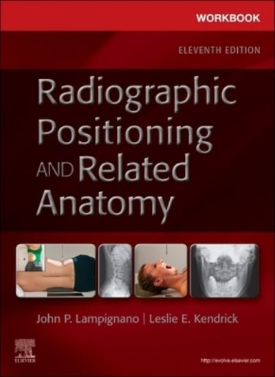
Workbook for Radiographic Positioning and Related Anatomy, 11th Edition

ISBN: 9780323936156
ISBN: 9780323936156
Εκδόσεις:
Διαστάσεις 28 × 22 cm
Μορφή

Εκδόσεις

Ημ. Έκδοσης

Σελίδες

Έκδοση

Κύριος Συγγραφέας

,

Original price was: 121,00€.Η τρέχουσα τιμή είναι: 108,00€.(Περιλαμβάνεται ΦΠΑ 6%)

Διαθέσιμο - Προπαραγγελία|Διαθεσιμότητα: 11-14 ημέρες

Περιγραφή

Use this practical workbook to reinforce your understanding of radiographic positioning and procedures! With chapters corresponding to those in Textbook of Radiographic Positioning and Related Anatomy, 11th Edition, this workbook provides a wide variety of exercises to help you apply important positioning principles and critically evaluate images. Included are laboratory activities, situational questions, self-tests, and image critiques to review and reinforce what you have learned with the textbook. The perfect study tool, this workbook prepares you to succeed on credentialing exams and in clinical practice.
New to this edition
  • NEW! Updated content matches the revisions to Textbook of Radiographic Positioning and Related Anatomy, 11th Edition, ensuring that information reflects the profession’s evolving technology and clinical practice.
  • NEW! The latest ARRT content specifications and ASRT curriculum guidelines prepare you for certification exams and for clinical practice.
  • NEW! Stronger focus on computed and digital radiography prepares you for the ARRT® certification exam and for clinical success
Key Features
  • A wide variety of review exercises include questions on anatomy, select pathology, and clinical indications as well as a positioning critique and image evaluation questions.
  • Situational questions describe clinical scenarios and ask you to analyze and apply positioning criteria to specific examples.
  • Laboratory activities provide hands-on experience performing radiographs using phantoms, practicing positioning, and evaluating images.
  • Image critique questions describe an improperly positioned radiograph then ask what modifications need to be made to improve the image, preparing you to evaluate the quality of radiographs produced in the clinical setting.
  • Chapter objectives provide a checklist for completing the workbook activities.
  • Self-tests at the end of chapters help you assess your learning with multiple choice, labeling, short answer, matching, and true/false questions.
  • Answers to the review exercises are provided at the end of the workbook for immediate feedback.

Περιεχόμενα

1. Terminology, Imaging, and Positioning Principles
2. Chest
3. Abdomen
4. Upper Limb
5. Humerus and Shoulder Girdle
6. Lower Limb
7. Femur and Pelvic Girdle
8. Cervical and Thoracic Spine
9. Lumbar Spine, Sacrum, and Coccyx
10. Bony Thorax—Sternum and Ribs
11. Cranium, Facial Bones, and Paranasal Sinuses
12. Biliary Tract and Upper Gastrointestinal System
13. Lower Gastrointestinal System
14. Urinary System and Venipuncture
15. Trauma, Mobile, and Surgical Radiography
16. Pediatric Radiography
17. Angiography and Interventional Procedures
18. Computed Tomography
19. Special Radiographic Procedures
20. Diagnostic and Therapeutic Modalities
Answers to Review Questions